When learning a task the most useful thing is to have a great teacher….why?? To watch what you are doing and give you the best feedback on your performance.
In physiotherapy, we often have to teach exercises for muscles located deep within our bodies (our core muscles – abdominals, pelvic floor, diaphragm). These muscles have a stabilising role within our bodies and as such have a principal function of damping down unwanted movements. This, in turn, means they are hard to observe working in real time.
Ultrasound scanning is not a new phenomenon, we have been looking at babies in the womb and checking their health and development since the 1950s. It is only in the last 10 years that physiotherapists have started to utilise it in their clinical care.
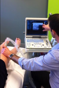
Real-time ultrasound is a tool that therapists use to give the best feedback as to whether patients are turning on the right muscles deep inside themselves.  It’s an important part of learning a new skill and enables the therapist to teach exercises more efficiently and accurately. It does not replace hard work on the part of the patient to perform the exercises but it can accelerate the outcome by ensuring that the skill acquisition phase occurs optimally.
More recently still, therapists are starting to learn how to perform musculoskeletal scans, to be able to observe tendinopathies, muscle tears, swelling within joints…and this is further helping them optimise the therapy intervention and refer on for more thorough investigations as needed.
All in all RTUS is a very valuable tool and in the hands of skilled and thoughtful therapists can be a tool that can assist in yielding optimal patient outcomes.
What is Real-time Ultrasound Imaging?
- Real-Time Ultrasound Imaging (RUSI) is a medical imaging technique that uses the echoes created by high-frequency sound waves to construct an image.
- A hand-held probe is placed on the skin and transmits 1-5 MHz sound waves into the body. When the sound waves hit a boundary between tissues of different densities (i.e./ between fluid and soft tissue, soft tissue and bone) some of the waves get reflected back to the probe. Other waves will continue on until they too reach a boundary that causes them to be reflected. The reflected waves are transmitted from the probe back to the ultrasound machine, and a two-dimensional image is constructed using the speed of sound in tissue and the time for each wave’s return.
Why choose RUSI?
- RUSI minimizes exposure to radiation, as it does not emit any potentially hazardous rays
- RUSI collects images during movement, so muscle activity can be assessed
Why use RUSI?
- OB/GYNs use RUSI to image unborn babies
- Sports physicians and radiologists use RUSI to locate muscle tears and to guide cortisone injections into a particular area
- Physiotherapists use RUSI to view the activity of deep stabilizing muscles
RUSI and Physiotherapy
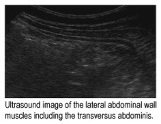
- The “core” or deep stabilizing muscles include the pelvic floor, transversus abdominus, multifidus, and diaphragm. Weakness in these muscles can often lead to low back pain, sacro-iliac pain, poor pelvic stability and overuse syndromes. Research also shows that after a back injury these muscles often are not functioning properly and need specific exercises to reactivate and strengthen them.
- RUSI allows the physiotherapist and the patient to view the activity in these muscles in real time. If the muscles are not activating automatically, RUSI can be used as a form of biofeedback to help the patient learn to selectively reactivate and strengthen these muscles. As the patient learns to use these core muscles, activation will eventually become automatic with movement.

