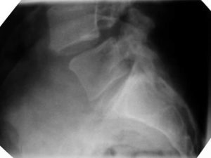
Spondylolisthesis occurs when one vertebra slips forward in relation to an adjacent vertebra.
In the x-ray above (the patient is facing to the left) you can see how the top vertebrae appears to be sliding off the front of the vertebrae below.
Isthmic spondylolisthesis is the most common form of spondylolisthesis. It has a reported prevalence of 5%-7% in the U.S. population and is the result of Spondylolysis (a defect in the pars interarticularis of a vertebra)
Spondylolysis is typically caused by a stress fracture of the pars which fails to heal and forms a chronic nonunion. It is especially common in adolescents who over train in activities such as tennis, diving, martial arts and gymnastics.
The next most common form of spondylolisthesis is degenerative, caused by arthritic changes to the spinal facets and disc. Long-standing segmental instability and gradual slippage occurs usually at the L4-5 level. This more commonly affects the older population.
A routine lateral (side) radiograph (see top picture) taken while standing confirms a diagnosis of a spondylolisthesis. The x-ray will show the translation (slip) of one vertebra over the adjacent level, usually the one below.
The Myerding grading system measures the percentage of vertebral slip forward over the body beneath
- Grade 1 is 0–25%
- Grade 2 is 25–50%
- Grade 3 is 50–75%
- Grade 4 is 75–100%
- Grade 5 is >100%
Symptoms
With Isthmic spondylolisthesis the presentation is often one of mild low back pain that occasionally radiates into the buttocks and posterior thighs, especially during high levels of activity. These symptoms can be attributable to the segmental instability present. Neurologic signs often correlate with the more advanced grades and can involve motor, sensory, and reflex changes corresponding to nerve root impingement
If the patient has complaints of pain, numbness, tingling or weakness in the legs, additional studies may be ordered. These symptoms could be caused by stenosis or narrowing of the space for the nerve roots to the legs. A CT or MRI scan can help identify compression of the nerves associated with spondylolisthesis.
The patient with degenerative spondylolisthesis is typically older and may present with back pain, radiculopathy (leg pain into the calf/foot with pins and needles/numbness and weakness), neurogenic claudication (increasing pain with walking, relieved by bending over), or a combination of these symptoms.
Treatment
With low grades of spondylolisthesis (either ishmic or degenerative) a conservative approach is warranted. This will involve physiotherapy aimed at improving segmental stability through motor control retraining of the deep muscle system. Here at SSOP we use state of the art Real Time Ultrasound to image these muscles to more effectively teach activation and control. Postural control exercises and stretches to tight muscle structures are also adjuncts to reducing pain and improving functional capacity. These are specifically targeted to your individual needs.
In the presence of neurological involvement, anti-inflammatory medication (NSAIDS) and/or injection therapy (under CT guidance) may be warranted to try to reduce the swelling around the nerve roots and reduce the compression on the neural tissue. This would be performed in conjunction with the above conservative measures (exercises).
Failure of these interventions to alter symptoms, or a progression of deteriorating symptoms may necessitate surgical fixation or fusion to stabilise the unstable segment artificially. This decision is usually made by a neuro or orthopaedic surgeon.
Most spondylolisthesis do not require surgical intervention however, and early diagnosis and management (including progression fro RTUS to clinical Pilates exercise programs) can assist in reducing pain and optimising your functional capacity.
For more info go to http://www.physioadvisor.com.au/8370650/spondylolisthesis-lower-back-injuries-physioad.htm


